P
PrintablesPaweł Ozga

Aorta
Segmentation of the aorta based on MRI + contrast, in the arterial phase. Reconstruction performed for preoperative preparation to correct aortic anatomy.
- Pobrania
- 138
- Polubienia
- 4
- Ocena
- 0
6 modeli
Segmentation of the aorta based on MRI + contrast, in the arterial phase. Reconstruction performed for preoperative preparation to correct aortic anatomy.
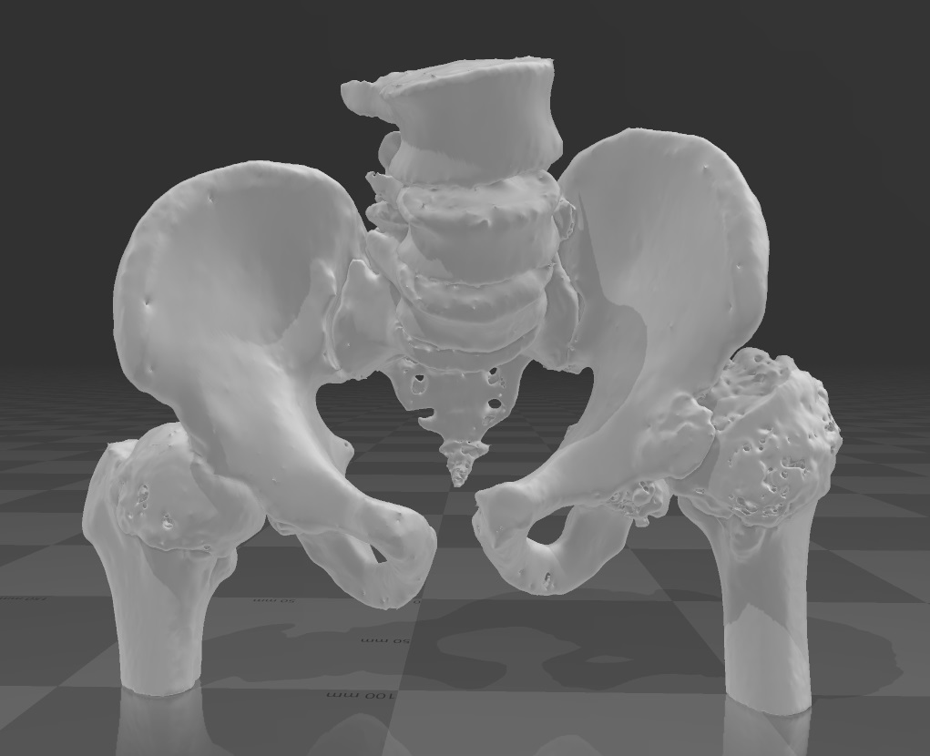
Reconstruction was based on CT scans, the purpose of the reconstruction was the evaluation of the progressive hip dysplasia and the fitting of the new implants: femoral head and acetabulum. The numerous "peeks" are errors resulting from…
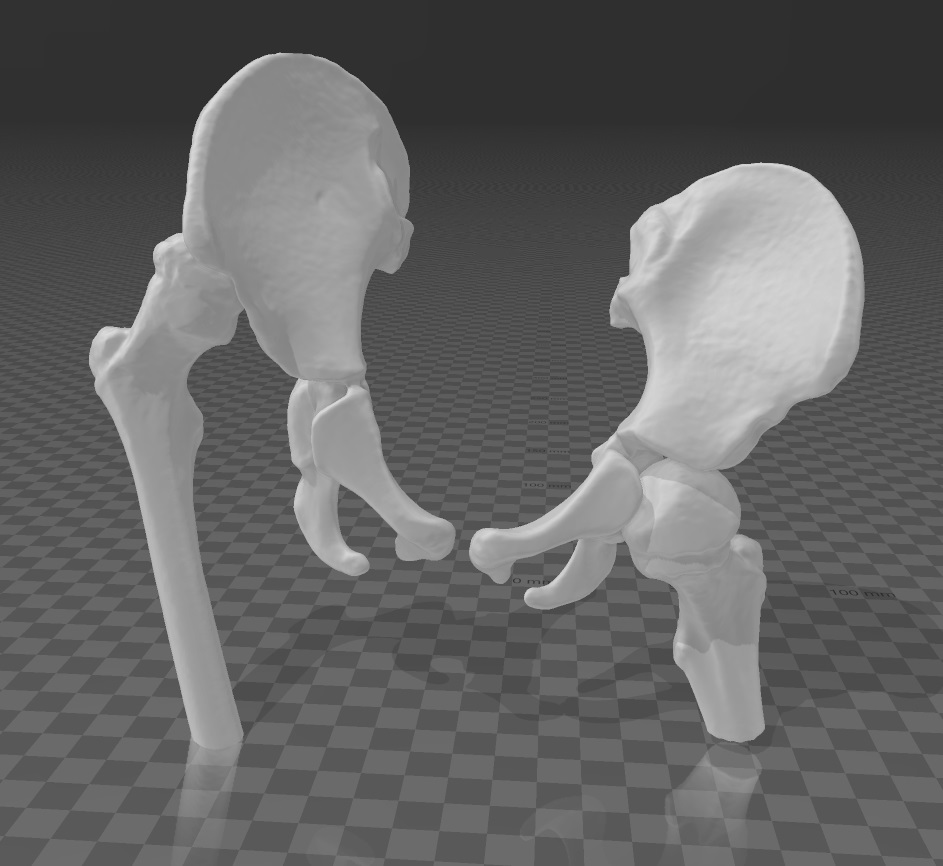
A pre-operative model of the hip joint including limbs, as I recall the child is male, aged about 2-3 years. Model prepared for University Children's Hospital of Cracow. Segmentation: MIMICS IS. What rarely happens, the quality of CT…
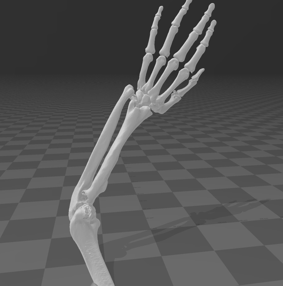
Cancer of the elbow joint, holes in the bone are the result of progressive degeneration of bone tissue resulting in a lack of X-ray absorption. The model was used to assess the pathology and plan treatment without performing a resection…
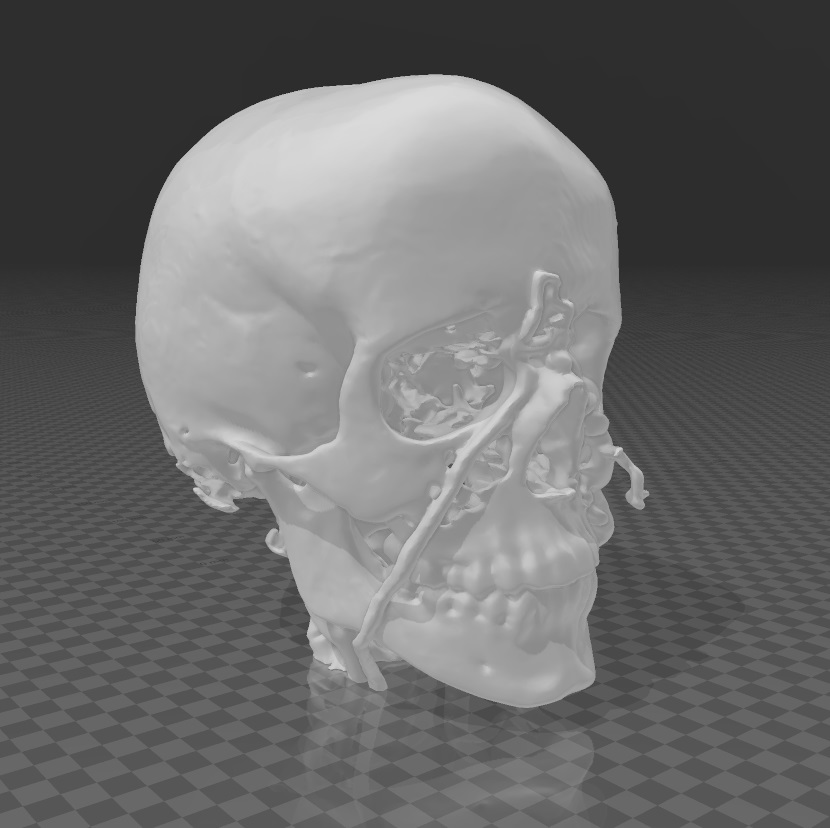
A very rare condition and a very sad story: as a result of the actions of the parents (yes both parents) and caregivers, the child suffered a permanent injury to the spinal column by bending it at 90 degrees. In order to restore the…
The model was segmented based on the CT + contrast , the aim was to capture the arterial phase. Segmentation was done in MIMICS IS, for University Children’s Hospital of Cracow (Poland) Model originally printed on SLS printer.
To wszystkie aktualnie dostępne wyniki.