
Lactate dehydrogenase sliced in half with removable FAD and substrates
We calculate the final price from your selected file. You review and accept it before payment.
Saved on this device.
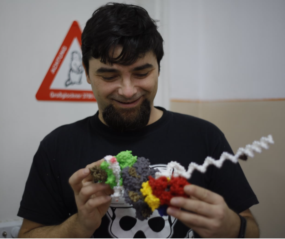
Surface model of lactate dehydrogenase (PDB ID 3LDH) showing the binding sites for NADH.
- Rating
- 0 / 5
- Likes
- 1
- Downloads
- 15
- Makes
- 0
- Views
- 276
- Reviews
- 0
Updated at source:
Model information
- Source
- Printables
- Category
- Chemistry & Biology
- License
- Creative Commons — Attribution
Surface model of lactate dehydrogenase (PDB ID 3LDH) showing the binding sites for NADH. Two models are provided: one for the complete homotetramer with 3 NAD's in place and one NAD binding site free; the second model is for a single homomer that was sliced in half in order to make the binding sites for NAD and pyruvic acid visible and accessible. The ligands are provided separated and can be inserted and removed. Model one can only be found in the zip archive and requires an IDEX printer for dual-color printing. Great for teaching protein/enzyme structure, as well as enzyme specificity. Made it to help me teach the biochemistry course.
Printer Technology/Material: Polylactic Acid (PLA)
Printer Make/Model: Creality Ender 5 Pro and Creality Ender 3 IDEX
Print Units: mm
Scale At Given Print Units: 200x and 100x
Pre- and Postprocessing Instructions
Printed at 0.2 resolution with 2 wall lines and 5% infill for the protein model; 0.2 resolution with 2 wall lines and 50% infill for the ligands. Supports are required. I have printed the homotetramer on an IDEX printer in order to highlight the NAD molecules ad the binding site. The model was oriented in such a way that after the printing of NAD was finished, the extruder 2 filament was changed to a different color just in time for printing the empty binding site.