
ACL Graft Tensioning Simulator with Digital Force Gauge – 3D-Printed Knee Trainer
Obliczymy ostateczną cenę na podstawie wybranego pliku. Sprawdzisz ją i zaakceptujesz przed płatnością.
Zapisane na tym urządzeniu.
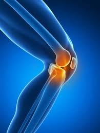
3D-printed knee simulator to train and measure ACL graft tensioning force with a digital force gauge.
- Ocena
- 0 / 5
- Polubienia
- 0
- Pobrania
- 2
- Wydruki
- 0
- Wyświetlenia
- 40
- Oceny
- 0
Aktualizacja u źródła:
Informacje o modelu
- Źródło
- Printables
- Kategoria
- Medical Tools
- Licencja
- Creative Commons — Attribution — Share Alike
🦵 What is this?
A 3D-printed simulator that reproduces the graft tensioning and fixation step of anterior cruciate ligament (ACL) reconstruction. A simulated graft (15 mm wide bovine leather strip) passes through the femoral and tibial tunnels of a CT-based knee model and is pulled from its tibial end, as during graft fixation. The other end is anchored to a digital force gauge, so all the force applied by the user is transmitted to the load cell and recorded in newtons. A display cover with a removable window allows blinded trials (display hidden) or training with visual feedback (display visible).
🔬 Validation status
This simulator was developed as part of a master's dissertation at the Graduate Program in Clinical Surgery, Universidade Federal do Paraná (UFPR), Brazil. Face validation and data collection with knee surgeons and medical students are ongoing (ethics approval CAAE 92701325.1.0000.0096). The published article will be linked here once available.
🎯 What is it for?
- Measuring how much force surgeons actually apply when tensioning an ACL graft, compared with how much they believe they apply.
- Assessing intra- and inter-individual variability of graft tensioning force across experience levels (specialists, residents and medical students).
- Training medical students to reach a target force using objective visual feedback from the force gauge.
🛠 How it was made
The femur, tibia and meniscus were modeled from computed tomography images, segmented and the simulator parts were designed in Autodesk Fusion 360. All parts were printed by fused deposition modeling (FDM) in PLA.
The main version was designed for the Instrutherm DD-2000 digital force gauge (push/pull, 196 N capacity, 0.02 N resolution, SD card datalogger). An alternative low-cost version is provided for the SHAHE AMF digital force gauge [model/capacity].
Instrutherm DD-2000 digital force gauge (link)
SHAHE AMF digital force gauge 200N (link)
📁 Files
STL files
- 01_Knee_with_meniscus – anatomical knee (femur, tibia, meniscus) with tunnels
- 02_Knee_base – base plate
- 03_Mounting_column – vertical column
- 04_Knee_shell_with_portals – external knee shell with arthroscopic portals
- 05_Force_gauge_holder_DD-2000 – holder for the Instrutherm DD-2000
- 06_Display_cover – cover that hides the gauge display
- 07_Display_cover_window – removable window (closed = blinded, open = feedback)
CAD files (Fusion 360 & STEP)
- Editable .f3d and .step files for all parts above
Low-cost force gauge option (SHAHE AMF)
- 08_Column_and_holder_SHAHE_AMF – .stl, .step and .f3z
🧰 Other materials needed
- Digital force gauge: Instrutherm DD-2000 (with its hook tip) or SHAHE AMF [model]
- Bovine leather strip, 15 mm wide, as the simulated graft
- Sutures at the tibial end of the graft for traction
- Screws/fasteners
- Optional: arthroscope and video monitor for arthroscopic viewing -
👥 Authors
Carolline Popovicz Nunes (master's student, validation study) – https://orcid.org/0000-0002-9835-0185
Lucas Santiago (master's student, simulator design) – https://orcid.org/0000-0001-7903-3996
Valentin Vanio Panatta Junior (medical student, simulator design) – https://orcid.org/0009-0006-0627-5162
Gabriela Pissetti Muniz (medical student, research assistant) – https://orcid.org/0009-0008-8088-1544
Edmar Stieven Filho (advisor) – https://orcid.org/0000-0003-2869-4019
Orthopedic Surgical Simulation Group, Hospital de Clínicas – Universidade Federal do Paraná (UFPR), and 3D Printing Laboratory, Centro de Simulação Profª Leide Parolin Marinoni, Curitiba, Brazil.
📌 📌 Zenodo DOI: https://doi.org/10.5281/zenodo.23020439
⚠ Disclaimer
For educational and training purposes only. Not a medical device and not intended for clinical use.